
DEEP:NEURO BrainSeg
DEEP:NEURO BrainSeg automatically segments key anatomical structures in brain MRI and provides regional volumes with age- and sex-matched percentiles to support quantitative assessment.
Product InquiryAutomated Brain Segmentation
Regional volume · reference percentile
MFDS Notified
No. D-26-92
PACS Integration
Review within your existing workflow
Quantitative analysis that complements brain MRI reading
BrainSeg automatically measures regional brain volumes and compares them against age- and sex-matched references, providing quantitative information that complements visual reading.
Regional volume measurement that complements visual reading
BrainSeg automatically measures regional brain volumes, helping quantify changes that are difficult to confirm by visual reading alone.
Volume comparison against age- and sex-matched references
Regional volumes are reported as percentiles against age- and sex-matched references, so you can see where each measurement stands.
Volume change versus prior studies
Regional volumes and change rates between prior and current studies are provided together, so changes over time can be confirmed.
Four core capabilities
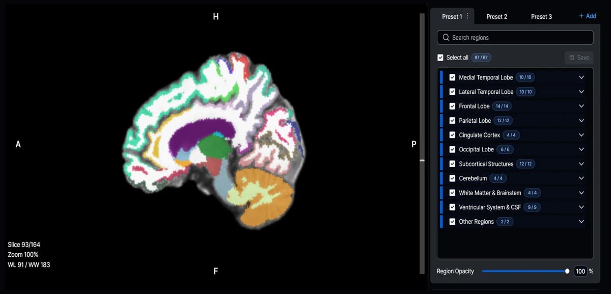
Automated segmentation and regional quantification
Automatically segments key anatomical structures in brain MRI and provides regional volumes with age- and sex-matched percentiles.
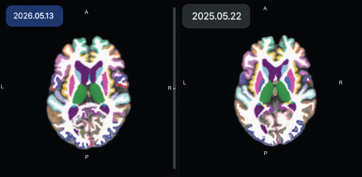
Longitudinal comparison for easy follow-up
Shows structure-level images and regional volume changes between prior and current studies on a single screen to support follow-up.
Automated Parkinsonism reference indices
Automatically measures the midbrain, pons, MCP, SCP, and ventricular structures to compute MRPI 1.0 and MRPI 2.0, providing quantitative reference information for differential assessment of Parkinsonian syndromes.
Integrated analysis report for efficient review
Delivers segmented brain MRI images and quantitative results in a single report, viewable within the existing reading environment through PACS integration.
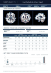
Automatically segments key anatomical structures in brain MRI and provides regional volumes with age- and sex-matched percentiles.
Patient communication that makes complex results easy to understand
Brain Age, 3D visualization, and patient-facing reports convey analysis results more intuitively and support result explanation and consultation.
3D visualization that shows results intuitively
Presents complex brain MRI analysis results as easy-to-understand images, helping communicate screening results effectively.
Brain Age that conveys complex quantitative data simply *
Presents brain imaging characteristics compared with the same age group as a Brain Age, conveying complex quantitative results intuitively.
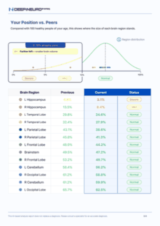
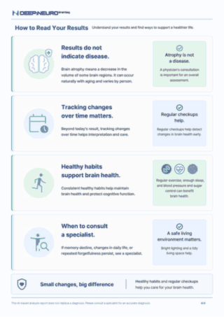
Personalized report showing results and trends together
Presents Brain Age and regional results in a patient-friendly format alongside trends versus prior studies, supporting ongoing follow-up and consultation.
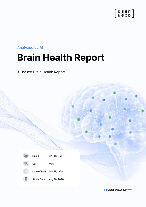
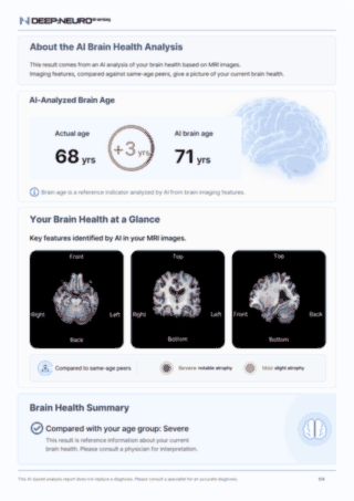
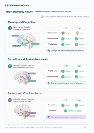
* Research Use Only
BrainSeg for reading, follow-up, and consultation
Radiology
Clinical needs
- Quantitative confirmation of regional brain volumes
- Volume change comparison with prior studies
- Analysis report review in PACS
How BrainSeg helps
Provides regional quantitative values and longitudinal changes to support brain MRI reading.
Neurology · Memory Clinic
Clinical needs
- Volume and percentile of regions of interest
- Volume change tracking across repeat studies
- Patient consultation using analysis results
How BrainSeg helps
Provides regional quantitative values and time-point changes to support follow-up assessment and patient communication.
Health Screening Center
Clinical needs
- Review of quantitative brain MRI results
- Result explanation using Brain Age
- Result delivery via examinee report
How BrainSeg helps
Brain Age and the examinee report help explain screening results more intuitively.
Clinical needs
- Quantitative confirmation of regional brain volumes
- Volume change comparison with prior studies
- Analysis report review in PACS
How BrainSeg helps
Provides regional quantitative values and longitudinal changes to support brain MRI reading.
Related Research
Through peer-reviewed publications and conference presentations, DEEP:NEURO continues its brain imaging analysis research.
DEEP:NEURO, expanding the scope of brain imaging analysis
Adopt DEEP:NEURO MRA and BrainSeg together to combine aneurysm detection support on brain MRA with quantitative volume analysis on brain MRI.
DEEP:NEURO MRA
Supports detection of unruptured intracranial aneurysms on brain MRA.
Learn moreDEEP:NEURO BrainSeg
Automatically measures regional volumes on brain MRI and provides quantitative information against age- and sex-matched references.
Current page